By Frangiska Mylona,
The term “inflammatory bowel disease” includes ulcerative colitis and Crohn’s disease. They are chronic inflammatory diseases of the intestines that follow a prolonged course with remissions and exacerbations, which last for years. Usually, they begin in adolescents and adults under 40 years of age. Both diseases can be associated with several extraintestinal manifestations like oral ulcers, pyoderma gangrenosum, and thromboembolic events, therefore a correct diagnosis can be difficult to make between these two, so one must be careful and precise.
Inflammatory bowel diseases can affect the same age groups with the same ratio between males and females. Still, it is been observed that Crohn’s disease affects chiefly the Jewish Ashkenazi populations. The exact etiology is still unknown to this day but both diseases , apart from their autoimmune profile , they share both environmental and genetic elements. As far as genetic predisposition is concerned, Crohn’s disease is associated with gene mutations in the innate immune system and autophagy-like mutations in the NOD2 gene. On the other hand, ulcerative colitis is associated with human leukocyte antigens (HLA) complex mutations .
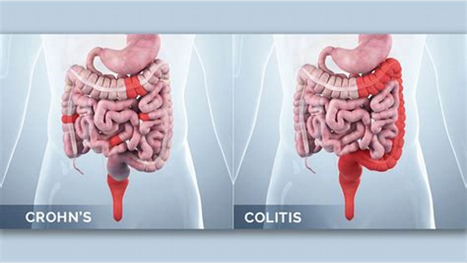
When it comes to the environmental factors, ulcerative colitis is most common in non-smokers or former smokers. It is interesting that patients undergone appendectomy or smokers are protected from developing ulcerative colitis . On the other hand , smoking is a factor that increases the risk for developing Crohn’s disease. Both diseases affect in a different manner the gastrointestinal tract. Crohn’s disease is a transmural process meaning it affects every part of the gastrointestinal tract leading to inflammation, ulceration, strictures, fistulas, and abscesses. The most common organs that get affected are the terminal ileum ,the ascending colon, the organs of the upper gastrointestinal tract and there is also a possibility for the patient to develop perianal diseases. Crohn’s disease is characterized by right lower abdominal pain, fever, weight loss, and even diarrhea , where in Crohn’s disease, non-bloody diarrhea is more common than bloody diarrhea. Since , in this disease , the terminal ileum is affected , it is possible for the patient to present with anemia due to Vitamin B12 deficiency .

Ulcerative colitis is a chronic and recurring disease that affects only the colon, unlike Crohn’s disease which affects the whole gastrointestinal tract. It is characterized by diffuse mucosal inflammation, which results in friability, erosions, and ulcers. The most common inflammation points include the rectosigmoid region (proctosigmoiditis), the left-side of the colon , and as well as the whole colon. In most patients, periods of symptomatic flare-ups and remissions are common while some of them may develop an aggressive course of the disease which increases the risk of hospitalization and surgery. This condition is characterized by bloody diarrhea, fecal urgency, tenesmus and left lower abdominal cramps, which are relieved by defecation. In severe form of the disease, the patient may present with hypovolemia and hypoalbuminemia along with weight loss , fever and tachycardia.
Laboratory findings are a useful tool when it comes to diagnosing every disease. In inflammatory bowel diseases, there are important laboratory values which can aid the physician in making the correct diagnosis . Important indications include disturbances in the levels of serum albumin as well as abnormalities regarding the complete blood count showing an increased amount of leukocytes due to inflammation .Leukocytes are associated with the elevation of fetal calprotectin which is an excellent marker for diagnosing Crohn’s disease . C-reactive protein (CRP), a common marker used for inflammation, may or may not be increased depending on the patient’s status. Other tests include examination of stool specimens to exclude species that can mimic symptoms of inflammatory bowel disease like Clostridium difficile.
The incidence of anemia due to iron deficiency or vitamin B12 deficiency, along with abnormal hemoglobin levels, is a common finding in Crohn’s disease due to the fact that patients suffer from malabsorption by the infected small intestine. Regarding ulcerative colitis , other important tests include the stool frequency, the amount of blood in stools, the hematocrit levels and the CRP values which along with the endoscopy findings , will help the physician to categorize the severity of the disease. Other helpful tools in diagnosis of IBD include the radiological imaging . Colonoscopy and specifically ileocolonoscopy with biopsy is preferred during the diagnosis of Crohn’s disease . When colonoscopy is incomplete, CT and further imaging methods are preferred , for not only in diagnosing but also in completing the staging of the disease .

However CT is limited when there is a need to check for complications such as perforation or abscesses. Some signs that can implicate Crohn’s disease include ; “skip areas” where inflamed bowel regions are interposed between healthy bowel. Other findings include ; narrowing , irregularities and ulcerations of the terminal ileum along with small bowel dilatation . Further signs which can be visualized are ; separation of the loops of the bowel due to fatty infiltration of the mesentery , known as “proud loop” , the “string sign” reflecting the narrowing of the terminal ileum due to spasm and fibrosis as well as possible fistulae which can be visible between the ileum and colon and also to the skin , vagina and the urinary bladder.
The GOLD standard for diagnosing ulcerative colitis includes biopsy and colonoscopy . As for the radiological CT findings in ulcerative colitis , thickening of the bowel wall is visible with irregular narrowing of the bowel’s lumen and infiltration of the surrounding fat . However, this radiological image could be found in other causes of colitis , therefore clinical and medical history are of great importance.
References
- CURRENT Medical Diagnosis & Treatment , 2024, edited by Maxine A.Papadakis MD, Stephen J.McPhee MD,Michael W.Rabow MD, Kenneth R.McQuald MD. Associate editor Monica Gandhi MD,MPH. Sixty-third edition.
- Davidson, Παθολογίας; Γενικές αρχές και κλινική πράξη της Ιατρικής ,22η Έκδοση/ 5η Ελληνική Έκδοση, Επιστημονικές Εκδόσεις Παρισιάνου Α.Ε.
- Learning radiology , recognizing the basics , 5th Edition by William Herring , MD , FACR
- Ulcerative Colitis Imaging; Available here


